Categories: Featured Articles » Interesting Facts
Number of views: 31361
Comments on the article: 0
What is an ECG, EMG, EEG?
 An ECG is an electrocardiogram, a recording of electrical signals from the heart. The fact that a potential difference arises in the heart upon excitation was shown as early as 1856, during the Dubois-Reymond era. The experiment proving this was set by Kelliker and Müller exactly according to Galvani’s recipe: a nerve running to the frog’s foot was laid on an isolated heart, and this “living voltmeter” responded with a jerking of the paw to each heart beat.
With the advent of sensitive electrical measuring instruments, it has become possible to capture the electrical signals of a working heart by applying electrodes not directly to the heart muscle, but to the skin.
In 1887, for the first time, it was possible to register a human ECG in this way. This was done by the English scientist A. Waller using a capillary electrometer (the basis of this device was a thin capillary in which mercury was bordered by sulfuric acid. When current was passed through such a capillary, the surface tension at the boundary liquids changed and the meniscus shifted along the capillary.)
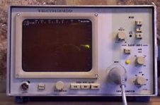
This device was inconvenient to use and the widespread use of electrocardiography began later, after the advent in 1903 of a more advanced device - the Einthoven string galvanometer. (The operation of this device is based on the movement of a conductor with a current in a magnetic field. The role of the conductor was played by a silvered quartz filament several micrometers in diameter, tightly stretched in a magnetic field. When a current was passed through this string, it bent slightly. These deviations were observed with a microscope. The device had low inertia and allowed to register fast electrical processes.)
After the appearance of this device in a number of laboratories, they began to study in detail how the ECG of a healthy heart and heart differs in different diseases. For these works V. Einthoven received the Nobel Prize in 1924, and the Soviet scientist A. F. Samoilov, who did a lot for the development of electrocardiography, received the Lenin Prize in 1930. As a result of the next step in the development of technology (the appearance of electronic amplifiers and recorders), electrocardiographs began to be used in every large hospital.
What is the nature of the ECG?
 When any nerve or muscle fiber is excited, current in some of its sections flows through the membrane into the fiber, and in others flows out. In this case, the current necessarily flows through the external environment surrounding the fiber, and creates a potential difference in this medium. This allows you to register the excitation of the fiber using extracellular electrodes, without penetrating into the cell.
The heart is a fairly powerful muscle. Many fibers are simultaneously excited in it, and a sufficiently strong current flows in the environment surrounding the heart, which even on the surface of the body creates potential differences of the order of 1 mV.
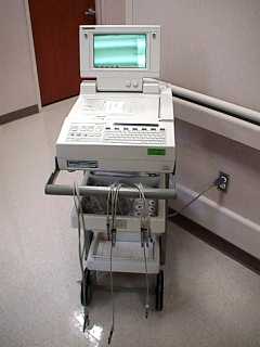
In order to learn more from the ECG about the state of the heart, doctors record many curves between different points of the body. To understand these curves, you need a lot of experience. With the advent of computer technology, it has become possible to significantly automate the process of "reading" ECG. A computer compares the ECG of a given patient with the samples stored in her memory and gives the doctor a presumptive diagnosis (or several possible diagnoses).
Now there are many other new approaches to the analysis of ECG. It seems very interesting. According to the data recorded from many points of the body, and their change in time, it is possible to calculate how the excitation wave moves through the heart and which parts of the heart become unexcited (for example, they are affected by a heart attack). These calculations are very laborious, but they became possible with the advent of computers.
Such an approach to ECG analysis was developed by L. I. Titomir, an employee of the Institute for Information Transmission Problems of the USSR Academy of Sciences.Instead of many curves that are difficult to understand, the computer draws on the screen the heart and the spread of excitation in its departments. You can directly see in which area of the heart the excitation is slower, which parts of the heart are not excited at all, etc.
The potentials of the heart were used in medicine not only for diagnosis, but also for controlling medical equipment. Imagine that a doctor needs to take x-rays of the heart at different phases of its cycle, i.e. at the time of maximum contraction, maximum relaxation, etc. This is necessary for some diseases. But how to catch the moment of greatest contraction? You have to take a lot of pictures in the hope that one of them will get into the right phase.
And so the Soviet scientists V, S. Gurfinkel, V. B, Malkin and M. L. Tsetlin decided to turn on the X-ray equipment from the ECG wave. This required a not-so-complicated electronic device, which included shooting with a given delay relative to the ECG wave. The witty solution of the problem in itself is especially interesting in that it was one of the first (now numerous) devices in which the body's natural potentials control certain artificial devices; This area of technology is called biofeedback.

The skeletal muscles of the body also generate potentials that can be detected from the skin surface. However, this requires more advanced equipment than for ECG recording. Individual muscle fibers usually work asynchronously, their signals overlapping each other are partially compensated, and as a result, lower potentials are obtained than in the case of an ECG.
The electrical activity of the skeletal muscle is called an electromyogram - EMG. For the first time, the potentials of human muscle fibers were discovered by listening to them using a telephone, the Russian scientist N. E. Vvedensky back in 1882.
In 1907, the German scientist G. Pieper used a string galvanometer for their objective registration. However, it was a complex and laborious method. Only after the cathode oscilloscope and electronic equipment appeared in 1923, electromyography began to develop intensively. Now it is widely used in science, medicine, sports, and also for biocontrol.
One of the first great uses of EMG biocontrol is to create prostheses for people who have lost their arm. Such prostheses were first created in our country.
And what is an EEG?
This is an electroencephalogram, i.e., the electrical activity of the brain, potential fluctuations created by the work of brain neurons and recorded directly from the surface of the head. Nerve cells, like muscle fibers, work simultaneously: when some of them create a positive potential on the surface of the skin, others create a negative one. Mutual compensation of potentials here is even stronger than in the case of EMG. As a result, the amplitude of the EEG is about a hundred times smaller than the ECG, therefore, their registration requires more sensitive equipment.
The EEG was first registered by the Russian scientist V, V. Pravdich-Nemsky on dogs using a string galvanometer. He introduced curare to dogs so that stronger muscle currents did not interfere with the registration of brain currents.
In 1924, the German psychiatrist G. Berger began at Jena University the study of human EEG. He described periodic fluctuations in brain potentials with a frequency of about 10 Hz, which are called the alpha rhythm. He first recorded the EEG of a person with a seizure of epilepsy and came to the conclusion that Galvani was right, suggesting that a section arises in the nervous system with epilepsy where currents are especially strong (cells there are continuously excited at high frequency).
Since we were talking about very weak potentials recorded by a little-known doctor, Berger's results did not attract attention for a long time; he himself published them only 5 years after the discovery. And only after in 1930they were confirmed by the famous English scientists Adrian and Matthews, they were "... stamped with academic approval", in the words of G. Walter, an English scientist who dealt with the clinical aspects of EEG in the laboratory of Gaul. In this laboratory, methods were developed that made it possible to determine the location of a tumor or hemorrhage in the brain by EEG, similar to how previously learned by ECG to determine the location of a heart attack in the heart.
 Subsequently, in addition to the alpha rhythm, other brain rhythms were discovered, in particular, rhythms associated with different types of sleep. There are a lot of EEG biofeedback projects. For example, if the driver constantly records the EEG, then you can use the computer to determine the moment, the code, he starts to doze, and wake him up. Unfortunately, all such projects are still difficult to implement, since the amplitude of the EEG is very small.
In addition to EEG - fluctuations in the potential of the brain in the absence of special influences, there is another form of brain potentials - called potentials (EP).
Evoked potentials are electrical reactions that occur in response to a flash of light, sound, etc. Since many neurons of the brain respond almost simultaneously to a bright flash of light, the evoked potentials are usually much larger than the EEG. It was no accident that they were discovered much earlier than the EEG (in 1875, by the Englishman Keton and independently of it in 1876 by the Russian researcher V. Ya. Danilevsky).
Using evoked potentials, one can solve interesting scientific problems. For example, after a flash of light, the response (EP) first occurs in the occipital region of the brain. From this we can conclude that it is in this region that signals about light come.
With electrical skin irritation, evoked potentials occur in the dark area of the brain.
With irritation of the skin of the hand, they occur in one place, the skin of the foot in another. You can map these answers and this map shows that the surface of the skin gives a projection onto the parietal region of the cortex of the human brain. It is interesting that in this design some proportions are violated, for example, the projection of the hand is disproportionately large. Yes, this is natural: the brain needs much more detailed information about the hand than, for example, about the back.
See also at bgv.electricianexp.com
:
